Tunnelled CVC Insertion
A Tunnelled Central Venous Catheter (eg Hickman Line) is a central venous access device (CVAD), most commonly used in Haematology patients for chemotherapy and transfusion of blood / blood components.
It is also the preferred CVAD for patients receiving long term nutrition directly into their bloodstream (eg TPN).
More about the Tunnelled CVC
It consists of a long flexible tube (catheter) which runs under the skin and enters either the large vein in the lower neck (internal jugular vein) or the vein under the collar bone (axillary vein). The other end of the catheter exits the skin on the chest wall several centimetres below the collarbone. This is where the nurses can access the line to take blood samples or administer drugs.
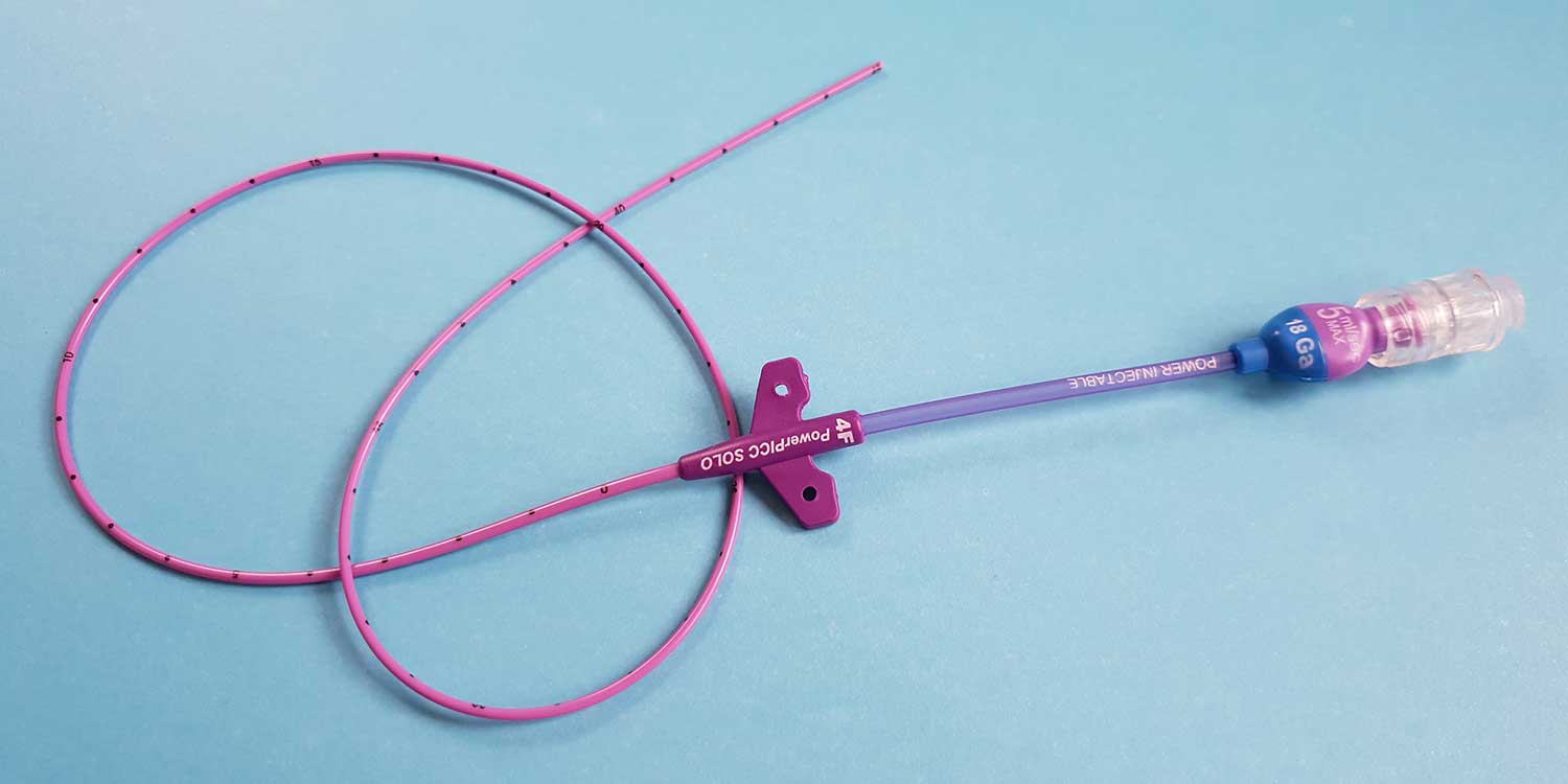
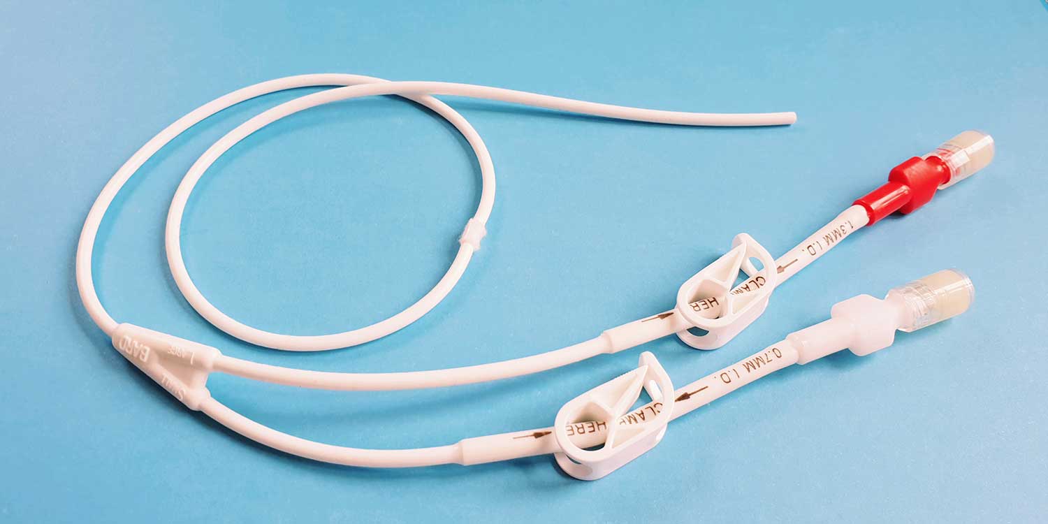
The main advantage of the Tunnelled CVC over the other CVADs is that it comes with either one, two or three lumens so multiple drugs or infusions can be given at the same time. The main disadvantage is that there is a long tube sticking out of the chest wall which can catch on clothing and be quite difficult to keep clean and dry.
How is the Tunnelled CVC inserted?
The procedure is performed under local anaesthetic and intravenous sedation in the Interventional Radiology suite at KIMS Hospital.
Dr Leech will examine the veins using an ultrasound machine to check their position and suitability and then insert a flexible wire into the vein. X-rays are used to check the final position of the line ensuring that the tip of the catheter lies in the large vein just outside the heart.
The actual procedure takes approximately 30 minutes. If the axillary vein is used, you will have a small incision (5mm) just under the collar bone and if the internal jugular vein is used this incision will be at the base of the neck. The catheter is then tunnelled and exits through the skin on the chest wall several centimetres below the collarbone (you will have two small scars in total). One or two strong sutures (blue in colour) are used to help anchor the line in place until your own tissue grows into the cuff which is fixed to the line. This cuff also helps to prevent infection.
Finally, the wounds are covered with water resistant dressings. Typically, most patients go home 30-60 minutes after the procedure. You should arrange for someone to take you home, as you must not drive for 24 hours following sedation.
What preparation is required?
All patients need to have MRSA swabs taken at least 48 hours prior to the procedure. If you are a Haematology patient or have already started chemotherapy you will also need to have some blood tests. If you are taking anticoagulant drugs (blood thinning medication such as Warfarin, Fragmin, Rivaroxaban), these will need to be stopped. Dr Leech will advise you when to stop your medication depending on the drug and the indication.
On the day of insertion, you may eat a light breakfast/snack before 9am if your procedure is in the afternoon but have nothing to eat after midnight if it is in the morning. You may drink water right up until the time of the procedure. You will need someone with you to drive you home afterwards because the procedure is performed under sedation and you must not drive or operate machinery for 24 hours.
Aftercare
For 3-4 weeks, avoid strenuous activities of the upper limb and chest wall and be very careful to keep the line dry when showering. You must not go swimming or soak in a bath the whole time the line is in place. If there are any issues associated with wound healing, persistent redness or soreness please contact your Oncologist, Chemotherapy nurse or Dr Leech’s secretary.
It is quite usual for your neck and chest to ache for a few days after the procedure. Simple pain killers such as paracetamol will help relieve this or you can take any other pain-relieving medication prescribed for you.
The blue anchor sutures will need to be removed after 10 days. This can either be done by the Chemotherapy nurses or by a practice nurse at your GP surgery.
The Tunnelled CVC needs to be flushed with saline once a week to keep it working and initially the dressings will also need changing every week. When the Tunnelled CVC is no longer required, it can be removed. This is normally done under local anaesthetic and is a more minor procedure than the insertion (sedation can be given if required).
Complications
With modern imaging techniques (using both ultrasound and X-rays), the risk of the procedure itself is minimal. However, there is still a very small risk of blood vessel injury, nerve damage, collapse of the lung and wound infection. There is also the very remote chance of being allergic to the local anaesthetic or the dressings that are used.
Bruising is relatively common especially in patients taking anticoagulant therapy or with low platelets but is normally confined to a small area and resolves within a couple of weeks. Thrombosis is a risk for all patients on chemotherapy and having a CVAD may increase this risk very slightly. If you do experience any sudden onset of pain and swelling in your arm, neck or face, you should contact your Oncologist, Chemotherapy nurse or Dr Leech’s secretary.
Rarely the catheter can move within the body and become malpositioned. This is known as migration and occurs more commonly with PICCs and tunnelled CVCs rather than ports. If a chest X-ray confirms that this has happened, it may be necessary to re-position the catheter.